OCULUS Keratograph 5M

Watch Video

OCULUS Keratograph 5M, yerleşik gerçek keratometreye ve harici görüntüleme için optimize edilmiş renkli bir kameraya sahip bir gelişmiş korneal topograftır. Özgün özelliklere meibomian bezlerinin incelenmesi, non-invazif gözyaşı filmi kırılma süresi ve gözyaşı menisküs yükseklik ölçümü ve lipid katmanı değerlendirmesi dâhildir.

The keratometer: integrated in all OCULUS topography devices

The benefits of the integrated keratometer are crucial for opticians’ and optometrists’ everyday practice:

  • measurements of actual corneal curvature
  • exact measurements for perfect correction of contact lenses
  • intuitive and simple to use
  • can be processed quickly
  • high-resolution images ensure clear results for customers

Functions

Measurements with Placido Ring Illumination

Thousands of measuring points are used to measure the whole surface of the cornea. A white ring illumination is used for this purpose. An infrared ring illumination is also provided for analysis of the tear film to prevent glare-related reflex secretion.

Measurements with Light Emitting Diodes

The perfect illumination has been integrated for every function of the Keratograph® 5M: White diodes for the tear film dynamics, blue diodes for fluo-images, infrared diodes for Meibography.

Software Highlights

Software

The heart of the Keratograph® 5M

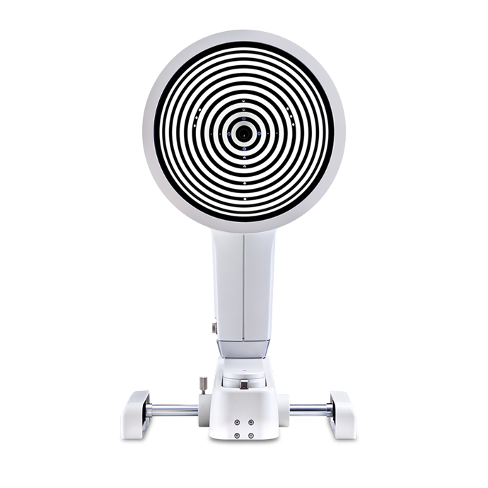
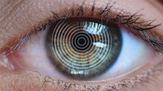
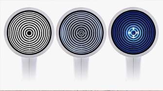
Makes the Tear Film Visible

The tear film is assessed using either white or infrared illumination. The new high-resolution color camera makes even the finest of structures visible. In addition to NIKBUT (Non-Invasive Keratograph® Break-Up Time) and measurement of the meniscus tear, the TF-Scan can also make an assessment of the lipid layer and the tear film dynamics. Tear film analysis with the OCULUS Keratograph® 5M is non-invasive and is conducted without any additional tools.

Make Dry Eye Diagnosis Crystal Clear

Find out the cause of dry eye disease quickly and reliably with the assistance of the new Crystal TEAR Report in the Keratograph® 5M. Carry out a comprehensive analysis using the measurement results as a basis for your diagnosis. All results are summarized in a patient-friendly, neatly arranged printout, enabling you to combine documentation and patient education.

How Does That Work?

The Imaging Software is used to record video and image files. In addition to viewing the videos and single images by themselves, you can also compare the recordings with the simulated fluorescein-images of the measured eye.

How Does That Work?

  • Simply integrate the Imaging Software into the Keratograph® software
  • Blue light-emitting diodes in the illumination beam path stimulate the fluorescein
  • Yellow filters are integrated into the observation beam path

Result: Static fluorescein-images and videos can be recorded under slit lamp conditions!

Oxygen Transmissibility of Soft Contact Lenses Made Visible

The OxiMap® presents a color map of the oxygen transmissibility of soft contact lenses based on the lens power, which is easy to understand – even for your customers!
An intact tear film and a good supply of oxygen to the cornea are an absolute necessity for wearing contact lenses comfortably. The OxiMap® presents a color map of the oxygen transmissibility of soft contact lenses based on the lens power – which even your customers will understand!

Indispensable for:

  • Fitting multi-focal contact lenses
  • Exact determination of the treatment area for refractive surgery
  • Seamless integration into the existing Keratograph® software
  • The infrared camera installed in the Keratograph delivers images of the
  • patient’s pupil, which are used as the basis for the measurements

Different Ways of Determining Pupil Reaction:

  • Examination of the pupil reaction both with and without glare
  • Examination using two different glare stimulus powers
  • Clear presentation of the results in graphic form: pupil changes over a period of time; minimum, maximum and mean pupil diameter, incl. standard deviation
  • Comparison views possible

Technical Specifications

Measuring range 0.1 – 1.5 in, 9 – 99 D
Accuracy ± 0.1 D
Reproducibility ± 0.1 D
Number of rings 22
Working distance 78-100 mm
Number of evaluated data points 22,000
Camera Digital CCD camera
Lightsource Placido illumination: White
Placido illumination: Infrared 880 nm
Fluorescein illumination: Blue 465 nm
Meibography: Infrared 840 nm
Tear film dynamics: White
Pupillometer illumination: Infrared 880 nm
Dimensions (W x D x H) 275 x 320 – 400 x 480 – 510 mm
Weight Measuring head: 7.1 lbs
With base: 13.5 lbs
Max. power consumption 18 W
Voltage 90-264 V AC
Frequency 47-63 Hz
Recommended computer specifications Intel® Core™ i5 (Current Generation), 1 TB HDD, 8 GB memory, Windows® 10 Pro
Recommended screen resolution 1920 x 1200 pixel