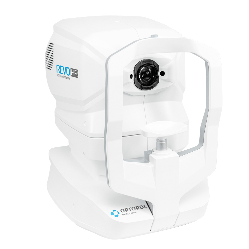
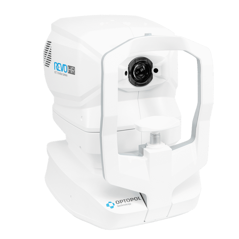
REVO HR
Spectral-Domain OCT | Color Fundus Camera
Watch VideoUltra High Resolution.
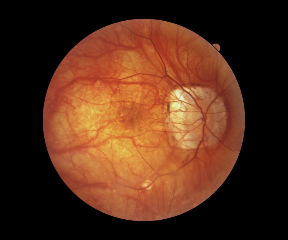
The REVO HR is an All in One device you can use in a number of ways such as a full color Fundus Camera or as a combo, providing simultaneous OCT and fundus images for high quality OCT imaging, including OCT-A.
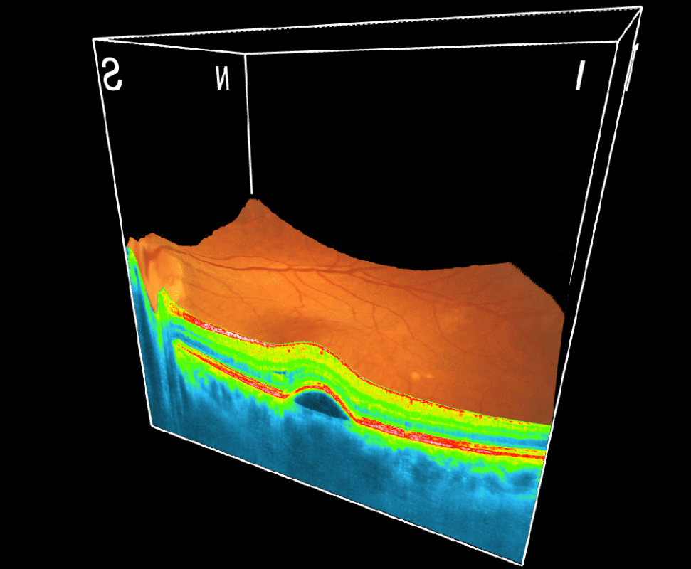
Ultra High Resolution
The combination of super-fast scanning at 130,000 scans per second and 3 µm High Resolution will provide a powerful tool for optimising precision, accuracy and improving the detection of the smallest lesions in tissue.
Spectral-Domain OCT
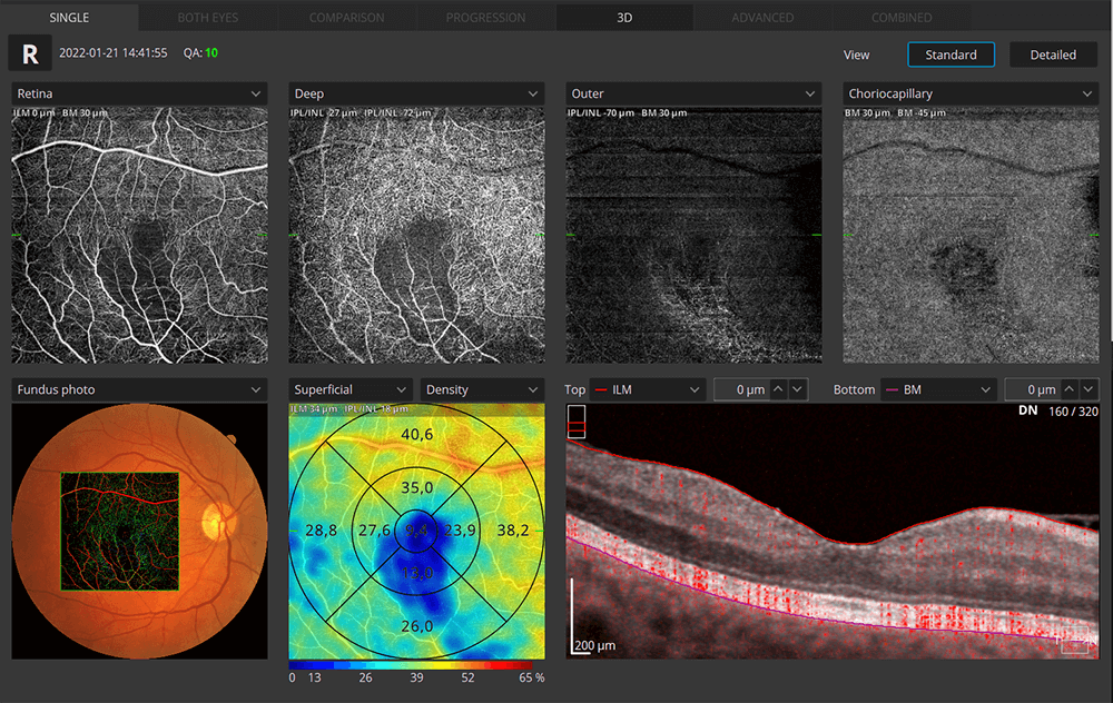
AI Segmentation

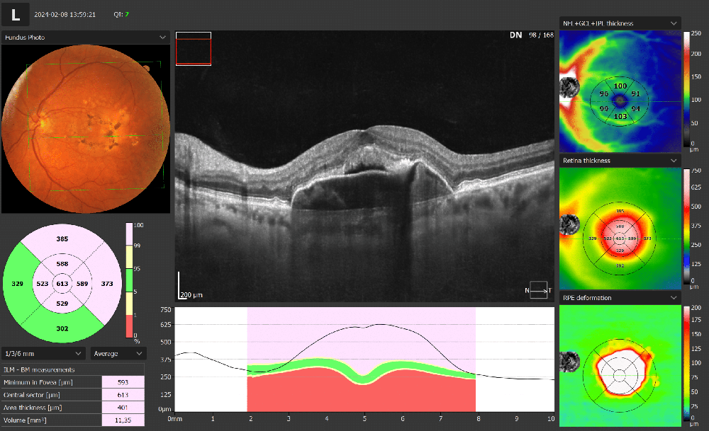
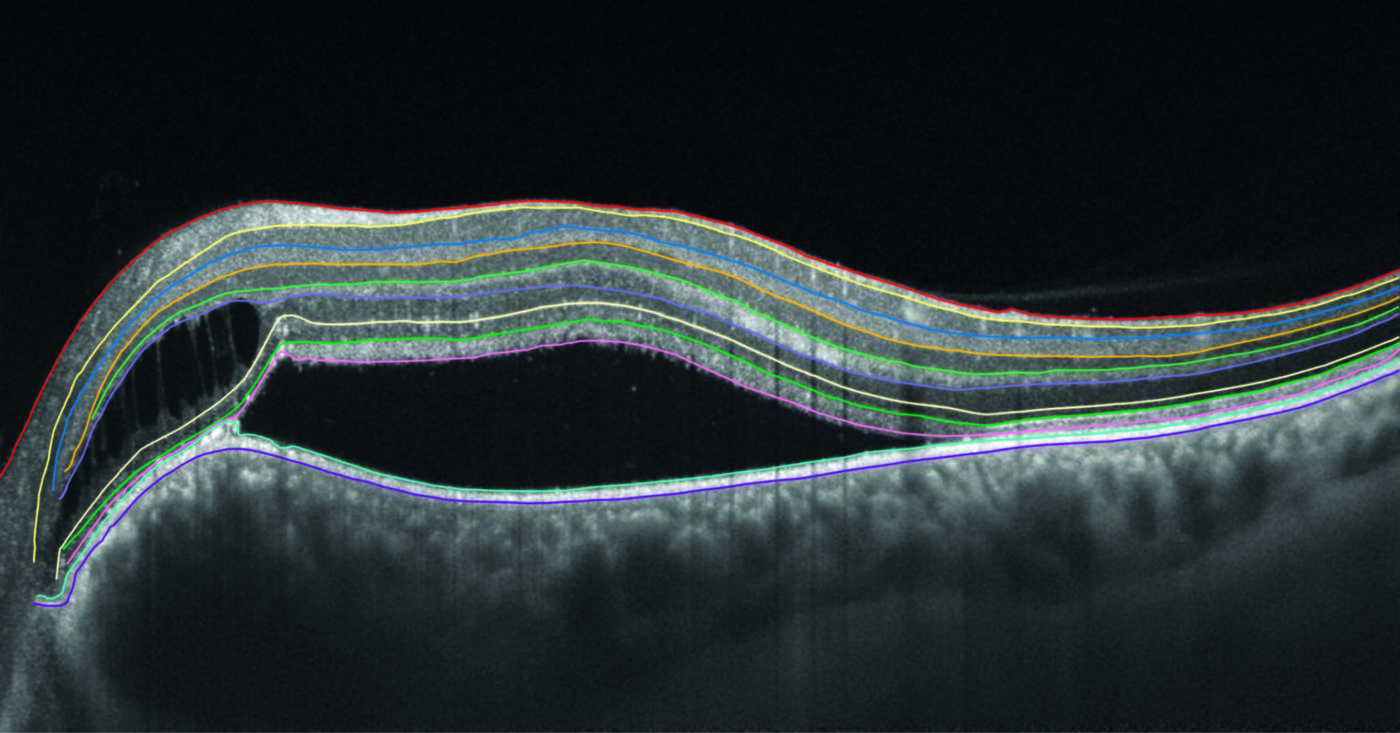
AI Retina
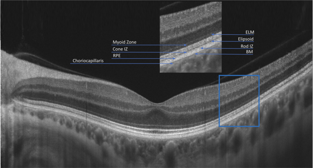
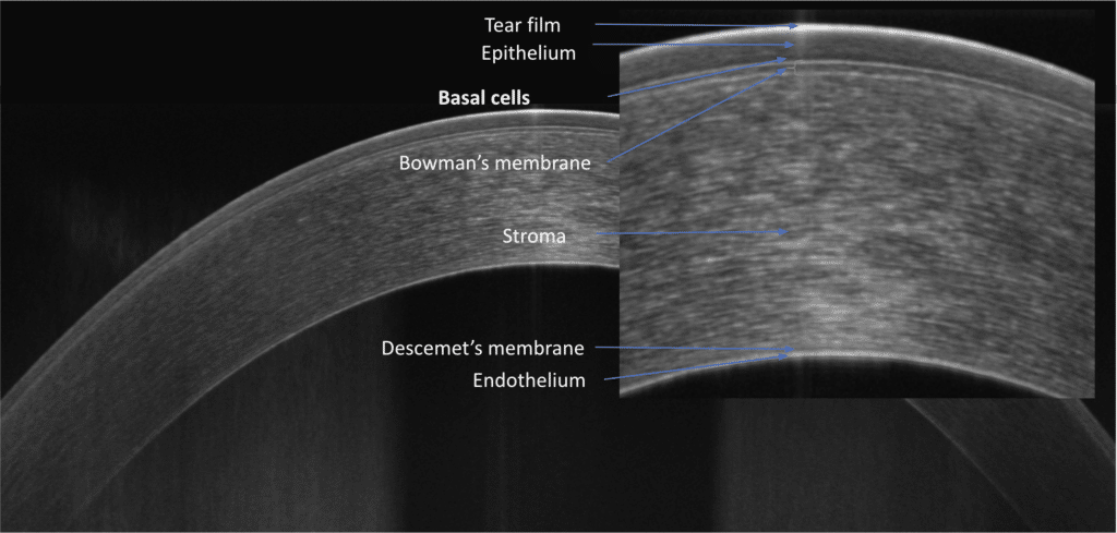
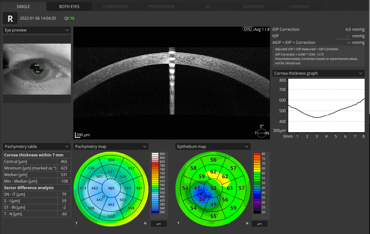
This new layer segmentation for the posterior segment is based on artificial intelligence, resulting in more accurate recognition of retinal layer boundaries. The AI system has a direct impact on the accuracy of the clinical assessment and the assessment of the status of areas of pathology in the retina Cornea and Epithelium Presentation of the results for both eyes allows quick and precise evaluation of the condition of the patient’s anterior segment. Epithelium and Pachymetry maps are included in the standard package.
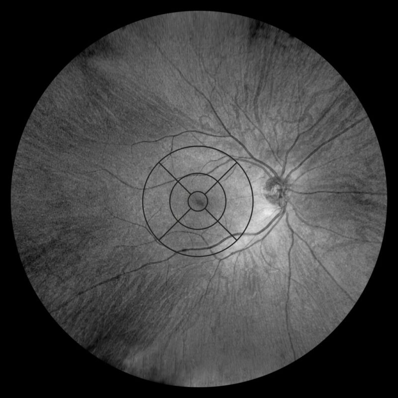
Widefield Analysis
A single Widefield 3D examination is now sufficient for the rapid assessment of both the retina and the glaucoma. Visualize and assess the thickness of the retina, ganglion cell, nerve fibers layers and optic nerve head on comprehensive data report when performing a rapid examination mapping up to 15×15 mm section. Widefield report presents horizontal and vertical tomograms and will include the topography of the disc creating helpful observation of glaucoma patients.
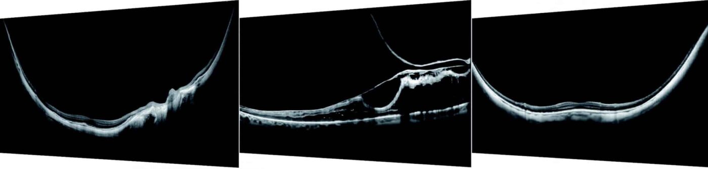
Cataract Mode
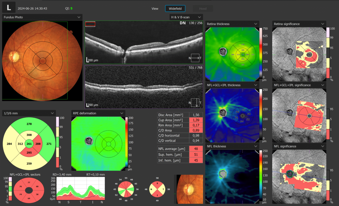
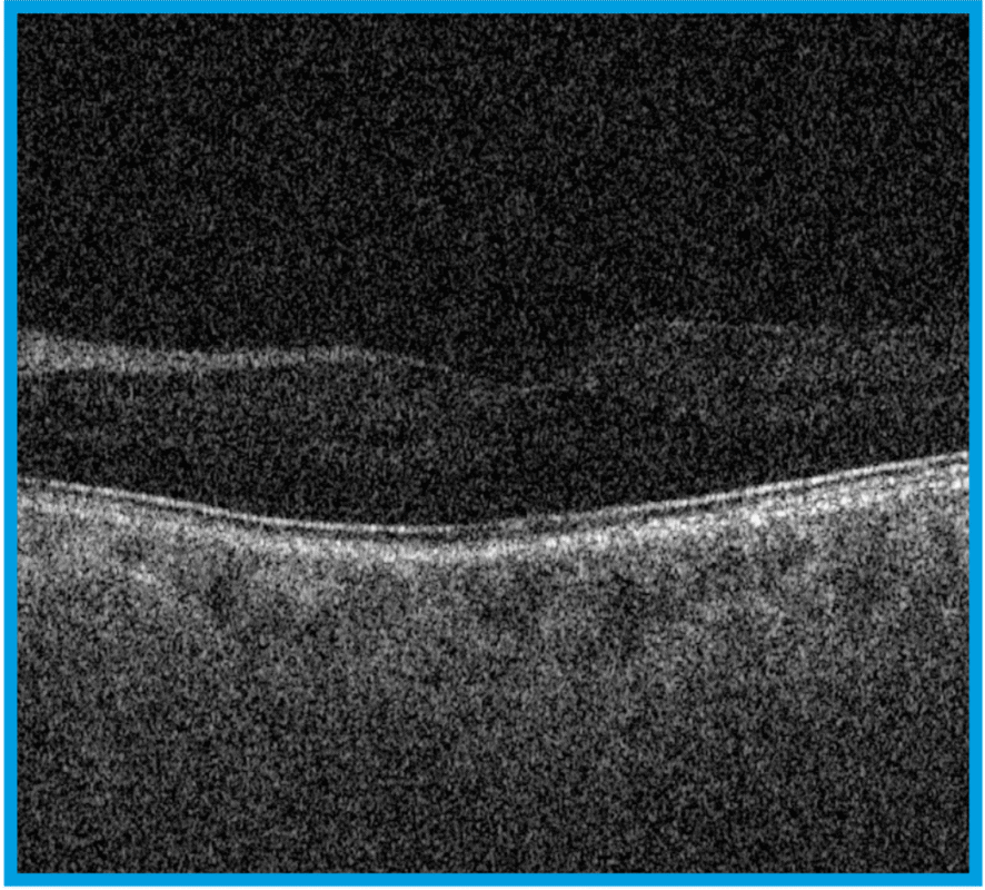
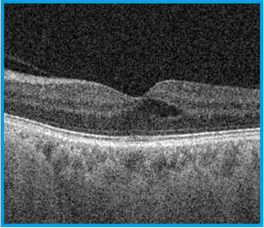
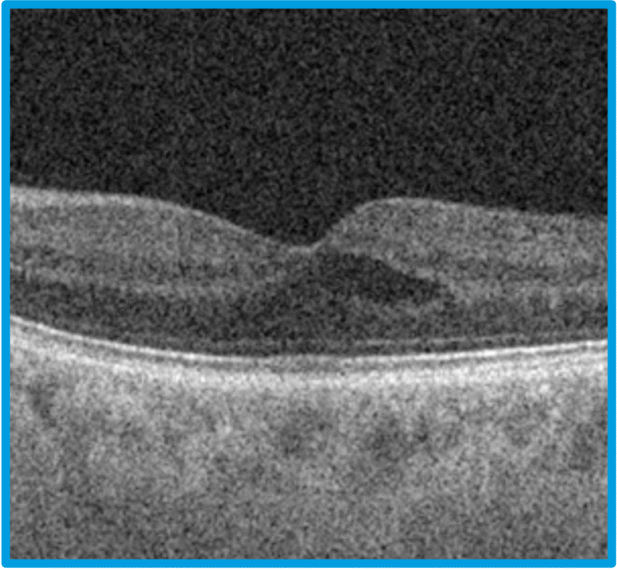
The cataract mode in the REVO series opens up new possibilities for patients with challening cases. This feature provides visualisation of structures hidden beneath opaque layers, making it ideal for diagnosing eye conditions that were previously difficult or impossible to study in patients with cataract, corneal oedemas or very dense floaters. The cataract mode allows the scanning speed and sensitivity of the OCT to be modified for better visualisation of patients with opaque media.
Normal – standard
scanning speed
Fine – balance
between speed and
scan saturation
Ultra Fine – max
sensitivity with most
saturated image
Maximum Intesity Projection
The MIP algorithm
Choose better visualization of angio data for analysis with the Maximum Intensity Projection (MIP) feature. This tool is useful for visualizing OCT-A data as it enables easier identification and tracking of high-intensity structures such as blood vessels.
AIP
MIP
Take your OCT to the next level
REVO HR

AccuTrack™
Our hardware-based eye tracker, compensates for blinks, loss of fixation and involuntary eye movements during scans reducing artifacts.

Auto Functions
Simplifying operation with the push of a button to auto-postion, auto-align, auto-focus, and auto-capture.

AI DeNoise
An advanced artificial intelligence (AI) algorithm removes noise from the tomogram for the highest image quality.

Custom Scan Protocols
Save time and never miss a scan. Create a custom preset group of scans and let the REVO capture all scans in order.

Motion Correction
The software-based motion correction (MC) compensates for involuntary eye movements and blinks by capturing two scans and generating a motion corrected scan when necessary.
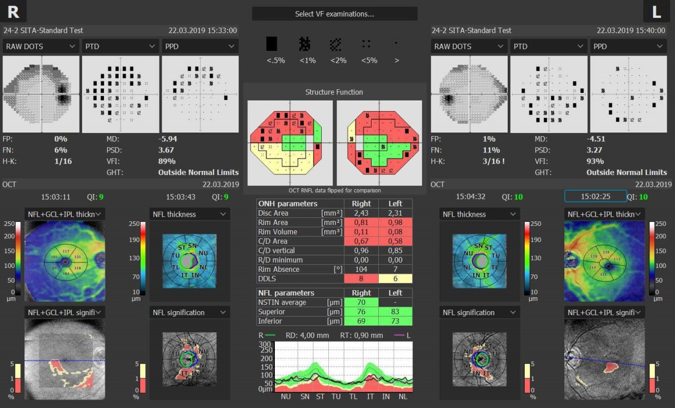
Structure + Function (S+F)
Comprehensive glaucoma solution that combines REVO OCT and PTS Visual Field results. S+F takes the diagnostic approach of the Hood report.
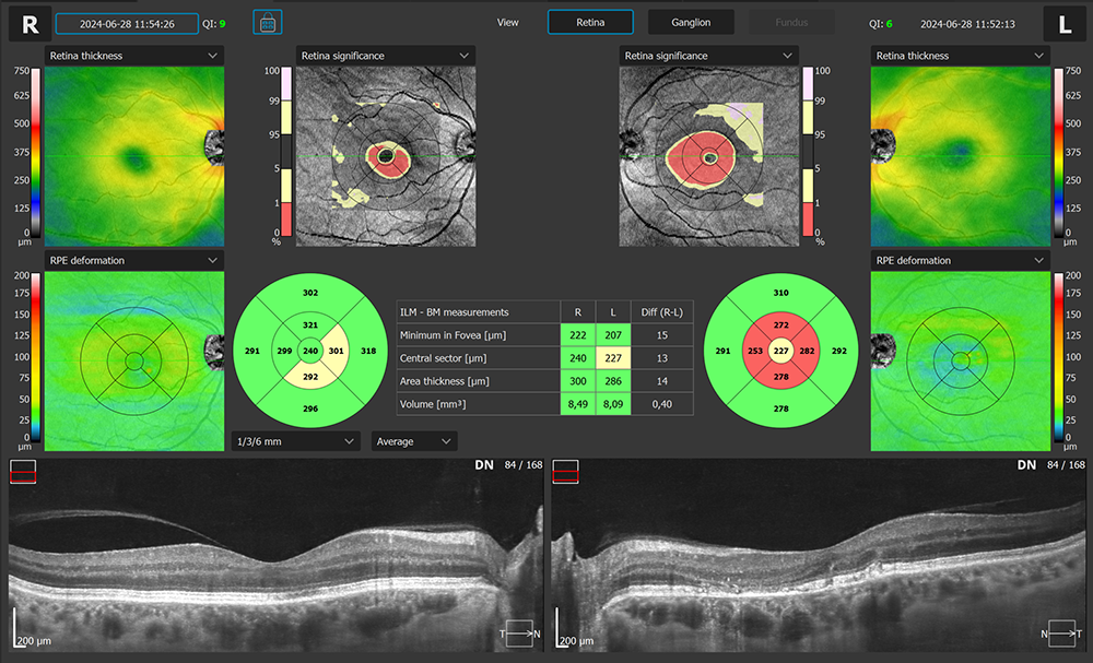
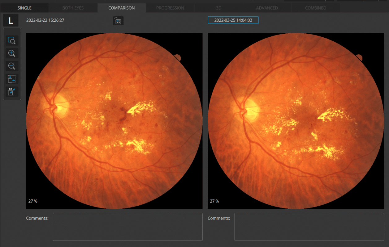
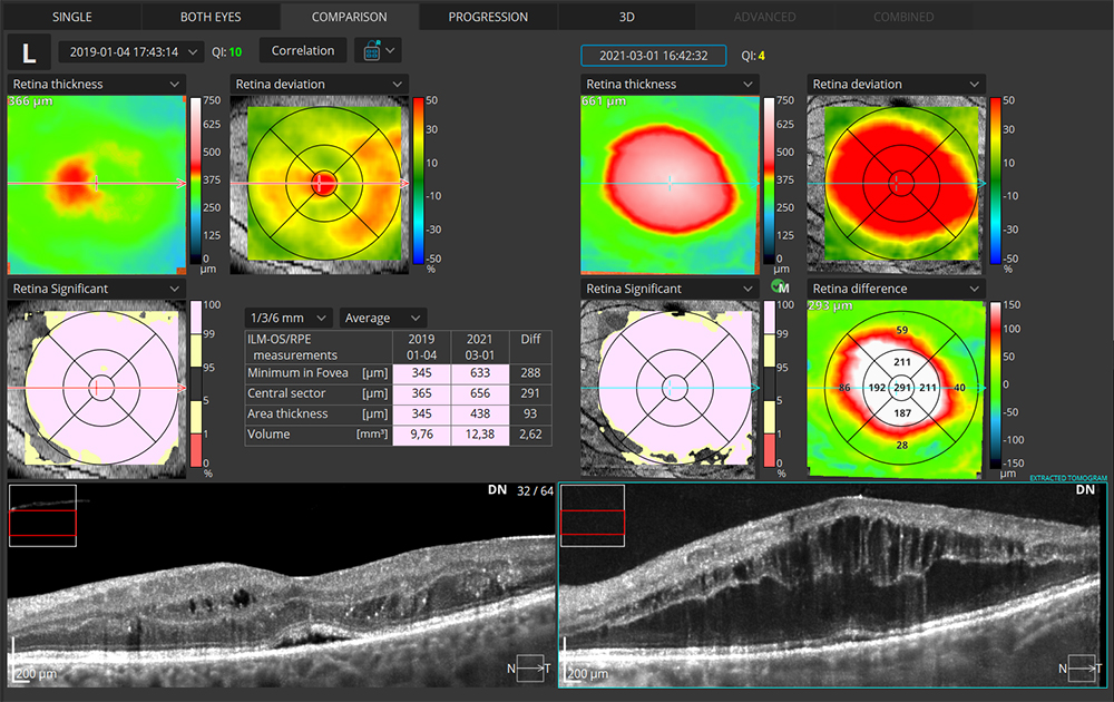
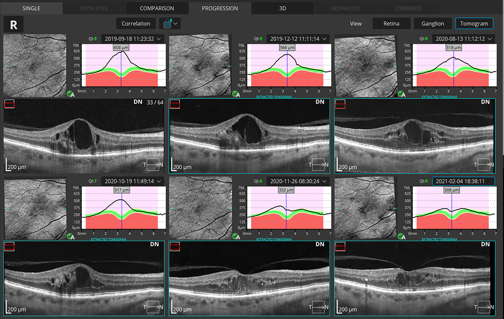
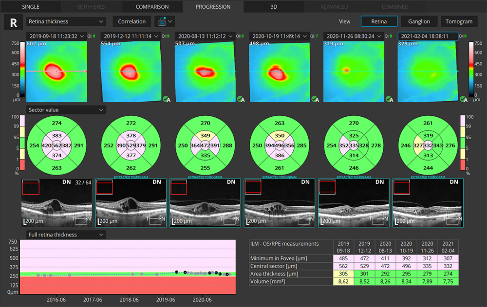
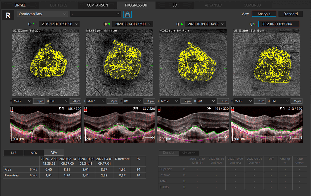
Progression Analysis
Gather baselines and follow-ups to monitor and manage disease progression in posterior and anterior scans.
Connectivity
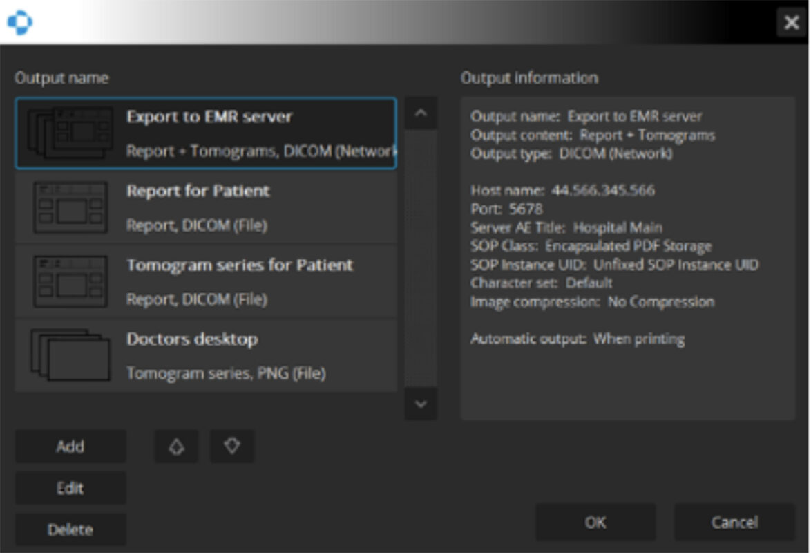
A proficient networking solution with DICOM and EMR capabilities. Quickly and easily export to a desired location.
Optional Software Modules
Follow disease progression
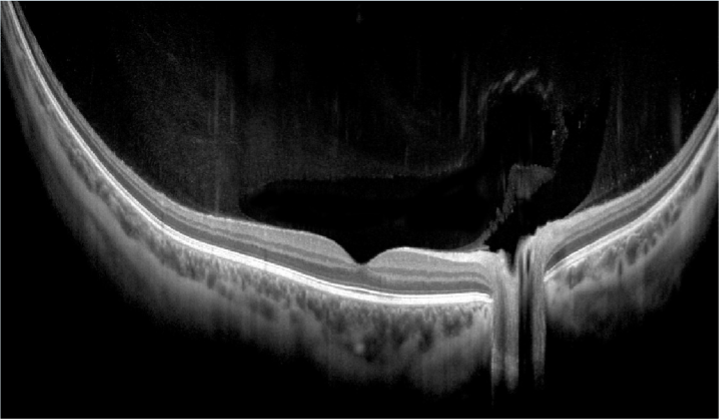
Full Range
With scans presenting New Extended DepthTM software, based on our Full Range technology, provides scans of increased depth for reliable and convenient observation of challenging cases. With scans presenting extended depth, this new imaging mode is perfect for diagnosing even highly myopic patients.
standardize scans performed
Connectivity
REVO offers an excellent networking solution that allows doctors to view and manage multiple examinations from review stations in your practice. The system comes with the latest version of Windows PRO and can easily export in PDF, JPEG and DICOM formats to your network or EHR.
Follow disease progression
Progression Analysis
Quickly view a chronological set of exams for analysis of changes in morphology, quantified progression maps, and progression trends.
standardize scans performed
Custom Scan Protocols
Save time and never miss a scan. Combine any scan type into a pre-set group. Choose a group of scans and set the order, the REVO will do the rest.
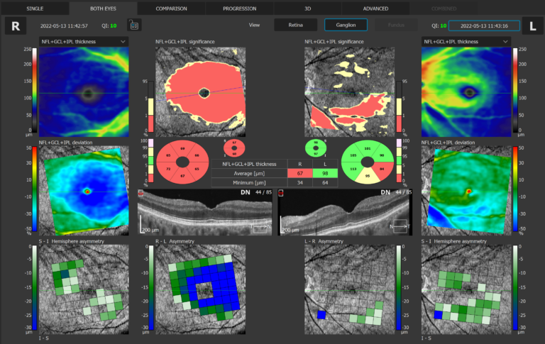
Ganglion Both
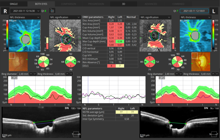
ONH Both
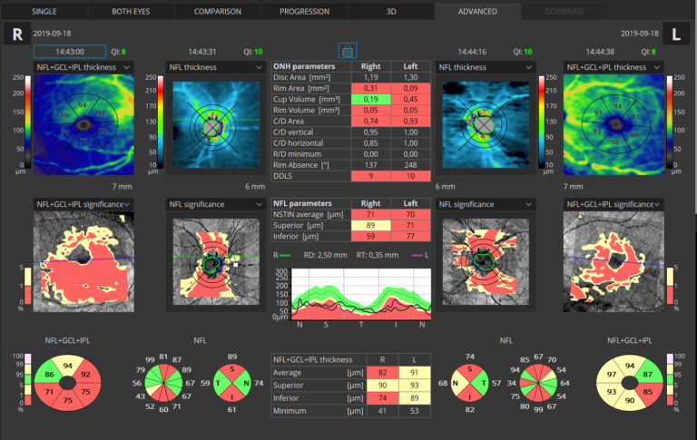
Advance Retina & ONH
ONH single
Follow disease progression
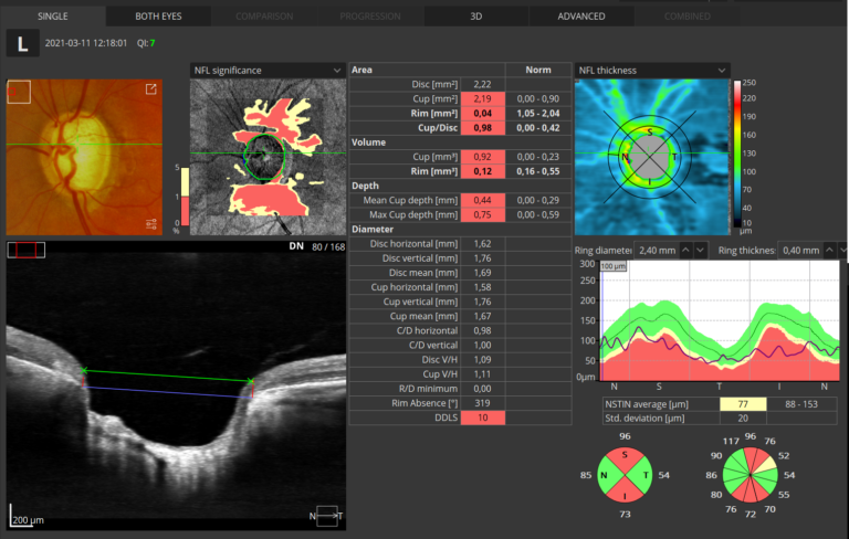
Glaucoma Tool Kit
Comprehensive glaucoma analytical tools for quantification of the Nerve Fiber Layer and Ganglion Cell Layer. The Disc Damage Likelihood Scale enables clinicians to precisely diagnose and monitor glaucoma for Optic Nerve Head analysis.
Comprehensive Hood report
Structure + Function
Our S+F report allows clinicians to understand the relationship between structural glaucoma damage and the functional impact on the patient’s field of vision. This provides a quick and comprehensive single-page report for glaucoma management.

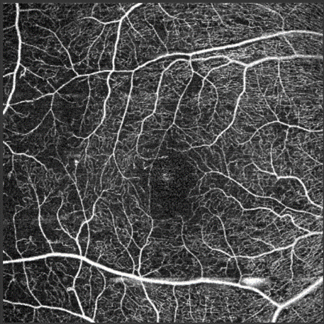
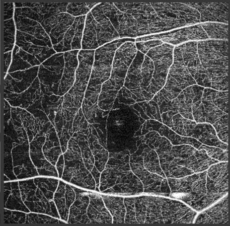
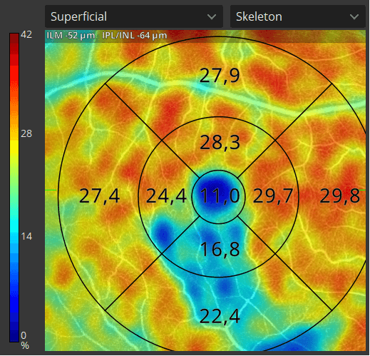
Angiography OCT
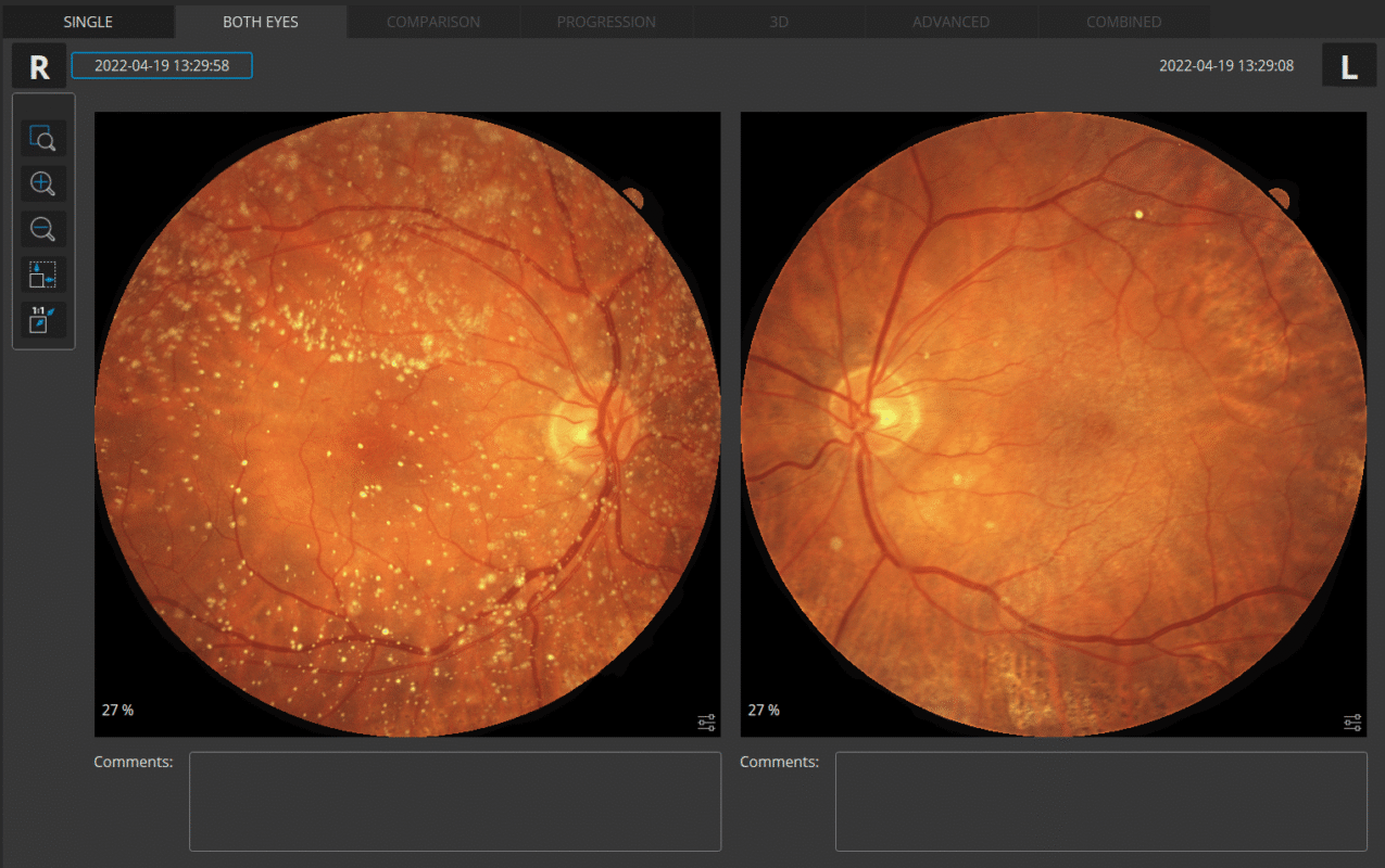
Angiography OCT provides an alternative to the traditional fluorescein method. Although OCT-A will not completely replace FA imaging, it is a quick and non-invasive tool. The software allows clinicians to observe, track and compare changes in the microvasculature of the retina in both eyes.
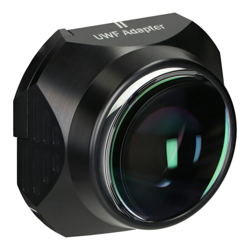
Ultra-Widefield Module
The UWF module provides a new wide perspective of imaging up to ~105º with a single scan. It allows the user to image the macular area along with the far periphery to capture the early stages of disease in the posterior part of the eye. The module allows 3D imaging for full analysis, averaging in enhancement mode, and angio OCT with the ability to visualise perfusion problems in the periphery.


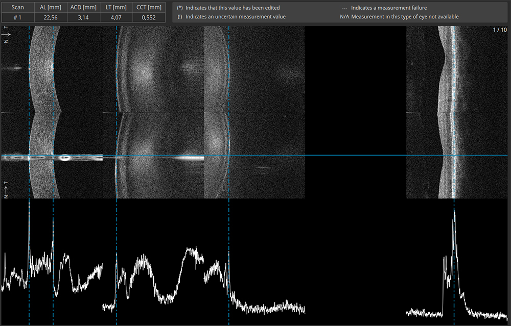
Biometry OCT
B-OCT® is an excellent tool for any clinician who manages myopia control or performs cataract surgery. Biometry OCT provides a complete set of ocular parameters: Axial Length, Central Cornea Thickness Anterior Chamber Depth, Lens Thickness, Pupil size, and White to White.

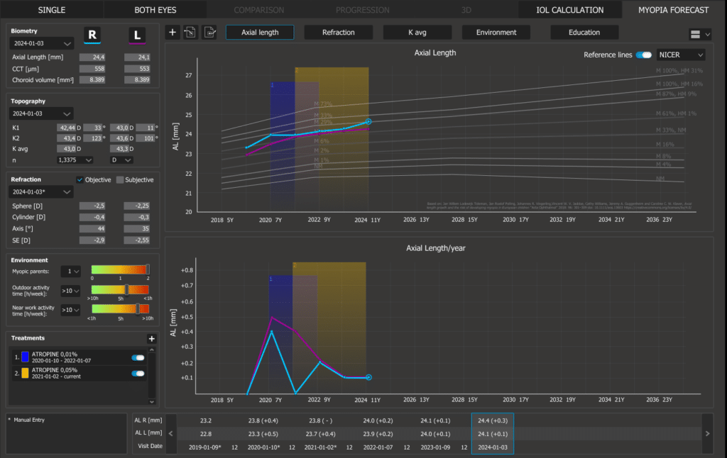
High Myopia
The Myopia Forecast module opens progression of the ocular structure parameters according to trends over population mode. Usage reference based on research from multiple universities along with environmental factors allow the monitoring of changes from childhood to adolescence.
The REVO offers an exclusive selection of reference data based on different studies over different time frames and demographics. Reference data can be selected from the NICER San Diez or Tideman studies.

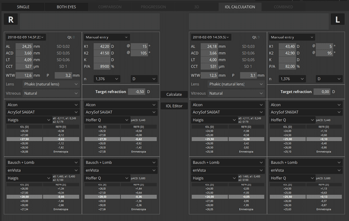
IOL Calculation
IOL formulas allow the user to calculate IOL implant parameters. Our systems now support the latest IOL data base standard IOLCon.org so that you can always keep your library up-to-date.

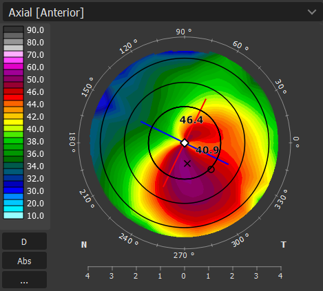
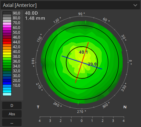
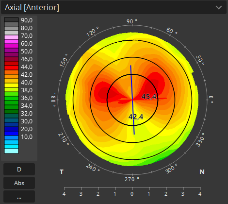
Topography
Topography OCT is a pioneering way to provide detailed corneal curvature maps. Anterior, Posterior surfaces and Corneal Thickness provide the True Net Curvature information. T-OCT™ is excellent when paired with the B-OCT® module for IOL surgery.