Advanced Imaging: The cornea and lens are displayed in a single image [Deep Imaging]
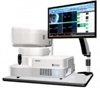
With CASIA2, light source coherence functions are improved, providing greater depth sensitivity compared to our previous model. Using this new technology, it is possible to measure from the anterior cornea to the posterior lens in a single shot. The scanning depth is approximately 13 mm. CASIA2 explores new possibilities in anterior segment analysis.
[Wide Imaging]
In corneal topography mode, it is possible to capture images around the angle. As with corneal shape analysis, it is possible to extract and analyze the angle and observe the IOL, enabling testing without switching measurement modes.
[Clearer Imaging]
Scanning 16 images simultaneously yields clearer images.
CASIA IOL Cataract Surgery (CICS) application for cataract surgery
CICS, the test application for cataract surgery, has been integrated into CASIA2, effectively supporting cataract surgery. There are two types in CICS: “Preoperative Test” and “Postoperative Test.” To use their functions, capture the image in each test protocol.
Performing Analysis Functions
Trend Analysis
In addition to the color-coded map showing parameter changes for each corneal shape, the simplicity of the chart means the information is intuitively easy to grasp. Therefore, this analysis is useful for observing keratoconus.
Lens Shape Analysis
By capturing the anterior cornea against the posterior lens, it is possible to measure the corneal curvature, thickness, and curvature of the anterior/posterior lens.