Wide measurement range and image capture of surrounding areas
Using unique technology, a large endothelial area of 0.25×0.54 mm can be imaged. The EM-4000 can measure a total of 13 points, including the central point, 6 peripheral locations, and 6 parafoveal locations, providing more options for imaging the endothelial area through corneal haze.
Automatic analysis and multi-screen functions
The built-in analysis software automatically displays 8 types of analysis values. The layout of the analysis results is selectable.
Captured images can be displayed in 4 ways (Photo / Trace / Area / Apex), allowing for a clearer view of the endothelium.
Dark-field analysis function
Automatically detects and excludes “dark areas,” such as the cornea guttata, from the analysis. This dark-field analysis is a unique function.
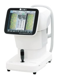
More convenient image capture at higher speeds
Display analysis results in approximately 4 seconds (2 seconds/eye) after measurement. Touch alignment is simple, and smooth and rapid image capture facilitates comfortable testing.
Internal database installed
A database is installed in the main unit. By displaying two patient datasets, comparisons can be made before and after surgery. We also provide a patient list screen for patient identification.