Topografik modellemenin öncüsü olan TMS’ye bir Scheimpflug kamera kurulur. TMS serisindeki en iyi cihaz. “Ring topo mode” ve “Slit mode” da ön/arka korneayı analiz etmek ve ön kamara ve lensi gözlemlemek mümkündür.
Topografik modellemenin öncüsü olan TMS’ye bir Scheimpflug kamera kurulur. TMS serisindeki en iyi cihaz. “Ring topo mode” ve “Slit mode” da ön/arka korneayı analiz etmek ve ön kamara ve lensi gözlemlemek mümkündür.
Scheimpflug camera for bright-light measurement
In addition to the functions of the previous TMS series, which projected corneal ring images, the TMS-5 can measure the anterior segment image (Scheimpflug image) by rotating and diffusing the slit light. The Scheimpflug camera’s slit mode uses a slit-cone method, making it less susceptible to external light interference, allowing for bright-light measurements.
*A removable headgear may be required depending on the test environment.
Anterior corneal map (fused) display to improve analysis efficiency
In cases of higher degrees of irregular astigmatism with a ring topo mode and slit mode measurement, the Scheimpflug image is combined with the ring topo data to provide an “anterior corneal fused map.” The Scheimpflug image covers the area not captured by the ring topo data, making it possible to display a fused map.
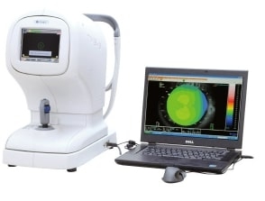
Instant capture and easy testing
A touch panel is integrated into the measuring head’s LCD screen, allowing basic operations to be performed from the main body. The joystick moves up and down, and the angle of the screen can be adjusted by the examiner.
Analyze all data from the anterior/posterior corneal segment with ring topography mode and slit mode.
4 map screens
If the Scheimpflug image is captured after ring topo data measurement with the ring cone, it is possible to create a combined anterior corneal map, an anterior/posterior corneal height map, and a pachymetry map.
Slit calculation screen: In the slit calculation screen, it is possible to observe the anterior segment and measure anterior chamber depth (ACD) and central corneal thickness (CCT).
[Map screen]1. Anterior corneal height mapEctasia scan
In the ectasia scan screen, both the anterior and posterior corneal shapes are assumed to represent the scan results for ectasia patterns, including keratoconus.
IOL power calculation software
“OKULIX” The IOL power calculation software “OKULIX” is available for verifying IOL power in cataract surgery after LASIK surgery.